All Departments
Emergency Cases
03328730300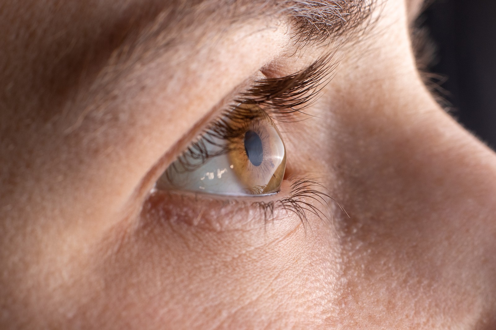
Keratoconus
WHAT IS KERATOCONUS?
Keratoconus (from Greek: kerato- horn, cornea; and konos cone) is a degenerative disorder of the eye in which structural changes within the cornea cause it to thin and change to a more conical shape than its normal gradual curve. Grossly the eye is more ‘cone’ shaped rather than the usual football or rugby ball shape.
Keratoconus can cause substantial distortion of vision, with multiple images, streaking and sensitivity to light. If afflicting both eyes, the deterioration in vision can affect the patient’s ability to drive a car or read normal print.
It is typically diagnosed in the adolescent years and attains its most severe state by the age of 30 (although this is variable)
Keratoconus affects around one person in a thousand. It seems to occur in populations throughout the world, although it is observed more frequently in certain ethnic groups, such as Asians. Environmental and genetic factors are considered possible causes, but the exact cause is uncertain. It is more common in patients with asthma, eczema and hayfever.
WHAT ARE THE STANDARD TREATMENTS FOR KERATOCONUS?
Some cases of keratoconus are mild and only result in the need for glasses. However, in most eyes the condition progresses and the cornea bows forward in an irregular manner. This irregular bowing of the cornea means that glasses or normal soft contact lenses are unable to give clear vision and such individuals may then require hard contact lenses in order to see. These hard lenses help the majority with keratoconus.
Other options include corneal ring segment inserts (Intacs or Ferrara rings), or topography-guided laser only to remove slight irregularity (not to remove prescription) and phakic implants. These are for people with stable keratoconus
However, keratoconus typically progresses up to the age of 30 necessitating changes in contact lenses and ultimately it is possible that contact lenses are no longer effective in restoring sight.
The only option is then to consider corneal transplantation (replacing the central cornea with donated corneal tissue). This is a major surgical procedure on the eye needing life long review with a risk of 1 in 500 of blindness and hence avoided if at all possible. There are risks transplant rejection, unpredictable visual outcomes, and development of high astigmatism. The advantages of postponing a transplant are many; even following a successful procedure, the mean survival rate of a penetrating transplant is about 17 years (many have to repeat the procedure).
CAN THE PROGRESSION OF KERATOCONUS BE STOPPED?
Yes. Cross-linking (also known as CXL, CCR, CCL and KXL) is a surgical treatment for keratoconus.
Corneal collagen cross-linking is a process whereby bonds form between collagen molecules within the structure of the cornea to stiffen its structure. Collagen is the main structural protein of the cornea. Corneal collagen cross-linkage occurs naturally with age and this process can be accelerated using a combination of riboflavin (vitamin B) eye drops and ultraviolet light shone at the cornea. It has been shown to strengthen the cornea. In keratoconus, it appears to prevent progression and may occasionally reverse the corneal bowing to a certain extent.
Cross linking was first developed in Germany in 2000 and clinical trials started the same year. In Italy, this has also been successfully performed since 2002. The procedure has been in routine use in Europe for a number of years with trials showing greater than 95% success rate. Results from an Australian study published in 2008 showed stabilization in all treated eyes, and a slight correction in visual acuity in most patients.
The aim is to stabilize the cornea and prevent progression of the keratoconus. In a small portion of people, there is a slight improvement in the shape of the cornea and hence the vision but this is not guaranteed.
WHAT IS THE PROCEDURE?
The 2 types of cross linking are epithelium on or epithelium off. At Midland Eye, we prefer epithelium off as we have had excellent results with this and do not want to take the chance of the treatment not working because the epithelium is on.
- Anaestheic drops are applied to the eye.
- The epithelium (the top layer of the cornea) is loosened and removed in the centre.
- A sticky yellow fluid, Riboflavin, is put on to this area for 20-30 minutes.
- The treatment is commenced for 30 minutes in 6 five minute steps. The patient needs to concentrate on a flashing light in the centre of the instrument.
- It is useful to bring an Ipod in as otherwise this is a long and boring procedure.
- The recovery is 3-5 days from the surgery. A stable situation is reached by 4-6 weeks.
WHAT ARE THE POSSIBLE RISKS AND COMPLICATIONS?
A slight, superficial haziness of the cornea has been seen in some patient in the first few weeks after treatment. This is transient and disappears by 6-8 weeks. It does not permanently impair vision.
Following treatment, it is possible to experience 24 to 72 hours of eye discomfort while the skin over-lying the corneal heals (the epithelium). Painkillers will be prescribed to alleviate this pain.
There is a risk of infection as with any operation. To prevent this potential complication, the procedure will be conducted under sterile conditions and antibiotics eye drops will be prescribed after the treatment.
Vision will be blurred for 1 weeks following the treatment and variable for a further month.
There is a risk of damage to other structures in the eye (the back of the cornea and lens) and hence we only do this procedure after we are happy that it is safe to do so following a full assessment.